
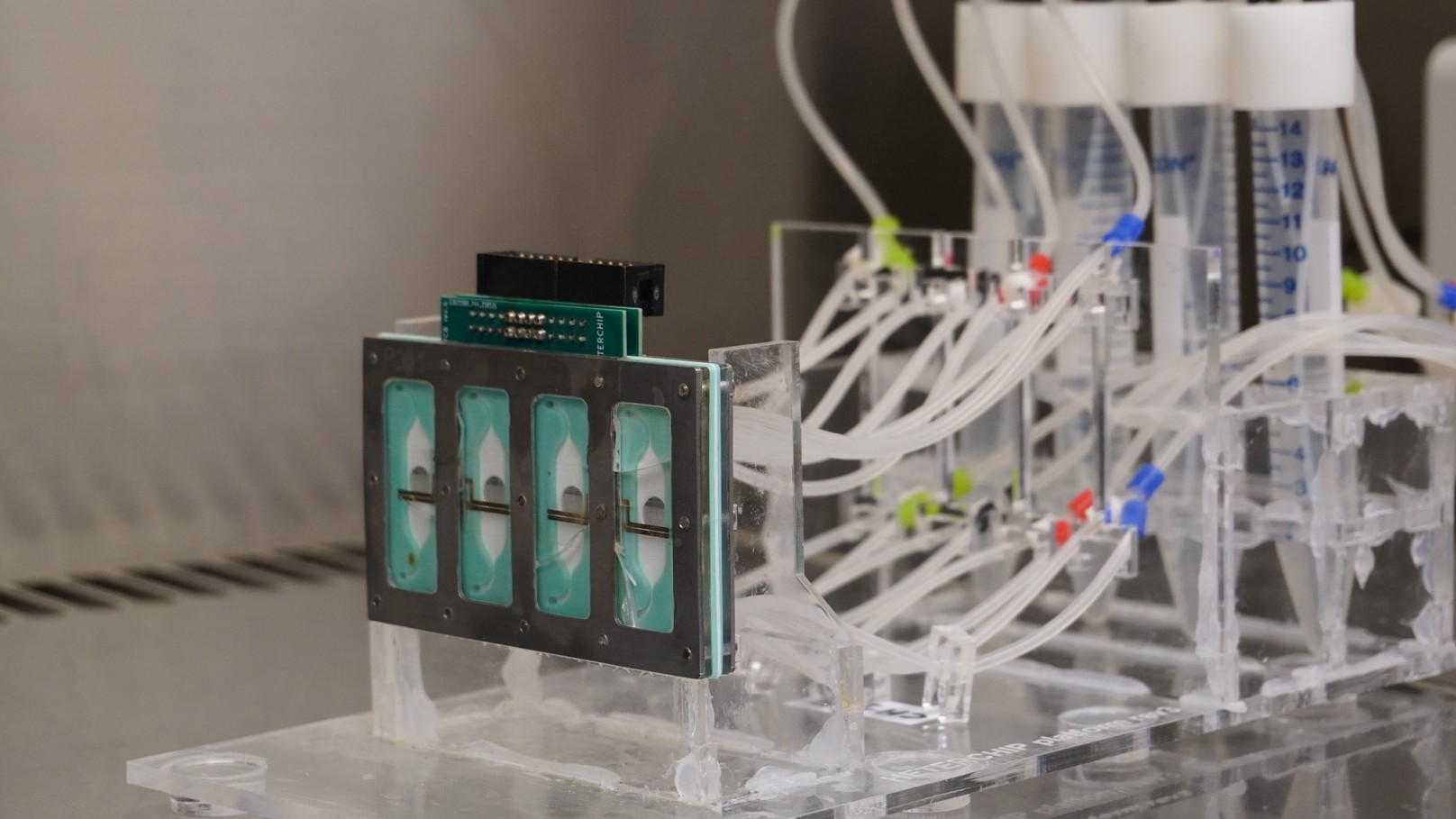
- Researchers from IDIBELL and IMB-CNM have developed a new vascularized cardiac model: a chip integrated into living heart tissue generated from human stem cells, which could advance our understanding of cardiotoxicity, one of the main barriers currently facing drug development.
- The new model is emerging as an alternative to reduce the use of animal models and avoid the rejection of promising drugs due to an overestimation of their cardiac toxicity.
The development of new drugs is a lengthy, costly process with a high failure rate: about 90% of drug candidates do not make it through clinical development. One of the main reasons is cardiotoxicity, the adverse effects that some drugs can have on the heart, which in many cases are not detected until advanced stages of clinical trials. Now, a team from IDIBELL’s Regenerative Medicine Program (RegenBell), led by Dr. Ángel Raya, together with the Barcelona Institute of Microelectronics (IMB-CNM-CSIC), has developed a “Heart-on-a-Chip”, a small microfluidic device capable of reproducing key aspects of the human heart function.
The research team presented the model in a recent study published in Advanced Healthcare Materials. The platform integrates three types of cardiac cells –cardiomyocytes, cardiac fibroblasts and endothelial cells– derived from the same stem cell line, enabling a more faithful recreation of the structure and behaviour of living cardiac tissue. Designed through bioengineering and manufactured entirely at IMB-CNM-CSIC, the chip could significantly improve the way drug safety is assessed before medicines reach patients.
A more accurate alternative to current models
Most experimental models currently used to study drug toxicity are based on cardiomyocytes –specialised heart muscle cells responsible for contraction– and expose the compounds directly to the tissue. However, this approach oversimplifies the complex organization of the heart by overlooking other cell types that are important for tissue architecture and by failing to reproduce how drugs actually reach the organ within the human body.
To overcome this limitation, the researchers have turned to organ-on-a-chip technology: microfluidic devices in which living cells are organised into perfused compartments or spaces recreating the physiological microenvironment. In essence, these systems replicate the function of human tissues on a miniature scale. In recent years, their potential as an alternative to animal models in biomedical research has attracted considerable interest.
“They have the potential to reduce animal experimentation in preclinical testing, but they can also improve the reliability of models, since they allow us to work with human cells,” says Dr. José Yeste, researcher at the IMB-CNM and co-author of the study. “Animal cells do not always respond in the same way as humans to certain compounds,” he adds. However, these technologies are still in the research phase and their industrial applications has not yet been fully established.
A miniature heart for drug testing
The team has developed a heart-on-a-chip consisting of three cellular layers: cardiomyocytes, cardiac fibroblasts –which provide structural support to the tissue– and endothelial cells –which line blood vessels (see Image 1). The three cell populations come from the same stem cell line, “which greatly reduces genetic variability and allows cellular interactions to resemble more closely those that actually occur in the human body,” explains Dr. Ángel Raya, coordinator of RegenBell and co-director of the study.
A key distinguishing feature of the model is the incorporation of endothelial cells. “By incorporating the endothelial layer, drugs are not administered directly onto the cardiac tissue, but must first cross the vascular layer, mimicking the way drugs reach the heart through the coronary circulation,” Raya adds.

Image 1. Three-layer cardiac cells composing the heart-on-a-chip: cardiomyocytes (green); cardiac fibroblasts (turquoise); and endothelial cells (red).
Regarding the technology itself, the IMB-CNM researcher and co-director of the study, Dr. Rosa Villa, clarifies that “the chip consists of a microfluidic channel system that enables the organised co-culture of different cell types, as well as control over the flow of nutrients and compounds.” This architecture facilitates the observation of cellular behaviour under dynamic conditions, more closely resembling those found in the physiological environment.
The endothelial layer: a protective barrier for the heart
To validate the model, the team studied its response to several drugs, such as doxorubicin, a widely used chemotherapy agent known for its cardiotoxic effects. The results revealed a significant difference between conventional models and the new three-layer system containing endothelial cells. In models lacking an endothelial layer, cardiomyocytes showed significant structural and functional deterioration. In contrast, in models incorporating endothelial cells, the cardiac tissue retained much of its contractile capacity and cellular organisation.
These findings suggest that endothelial cells play a protective role against certain toxic effects and that simplified experimental models may be overestimating the cardiac toxicity of some compounds by failing to include endothelium. According to the researchers, this limitation could contribute to the premature rejection of potentially valuable therapeutic candidates during the preclinical stages of drug development.
A further step towards advanced human tissue models
This work is part of IDIBELL’s strategic commitment to regenerative medicine and builds on the advances achieved by RegenBell in the development of increasingly complex and realistic human tissue models. Following the group’s recent achievement in generating cardiac tissue through 3D bioprinting for the first time (news), collaboration with the IMB-CNM -whose strategic priorities include promoting microtechnologies for biomedical applications- has now enabled the creation of a miniaturized, vascularised heart on a chip capable of reproducing more faithfully how the human heart responds to drugs.
The ultimate goal is to develop experimental systems that are increasingly closer to human physiology, helping researchers better understand disease, accelerate the development of new therapies, and progressively reduce the need for animal models in research through more accurate and clinically relevant preclinical platforms.
The development of this platform has been possible thanks to a close collaboration between IMB-CNM’s technological expertise and IDIBELL’s biological research capabilities. While the IMB-CNM has been responsible for the design and manufacture of the microfluidic device, the IDIBELL team has carried out the generation, integration and culture of the human cardiac cells. Both groups are members of the CIBER-BBN consortium (Center for Biomedical Research in Biomaterials, Nanomedicine and Bioengineering), a network that promotes interdisciplinary projects and was, in fact, were the origin of this line of research.